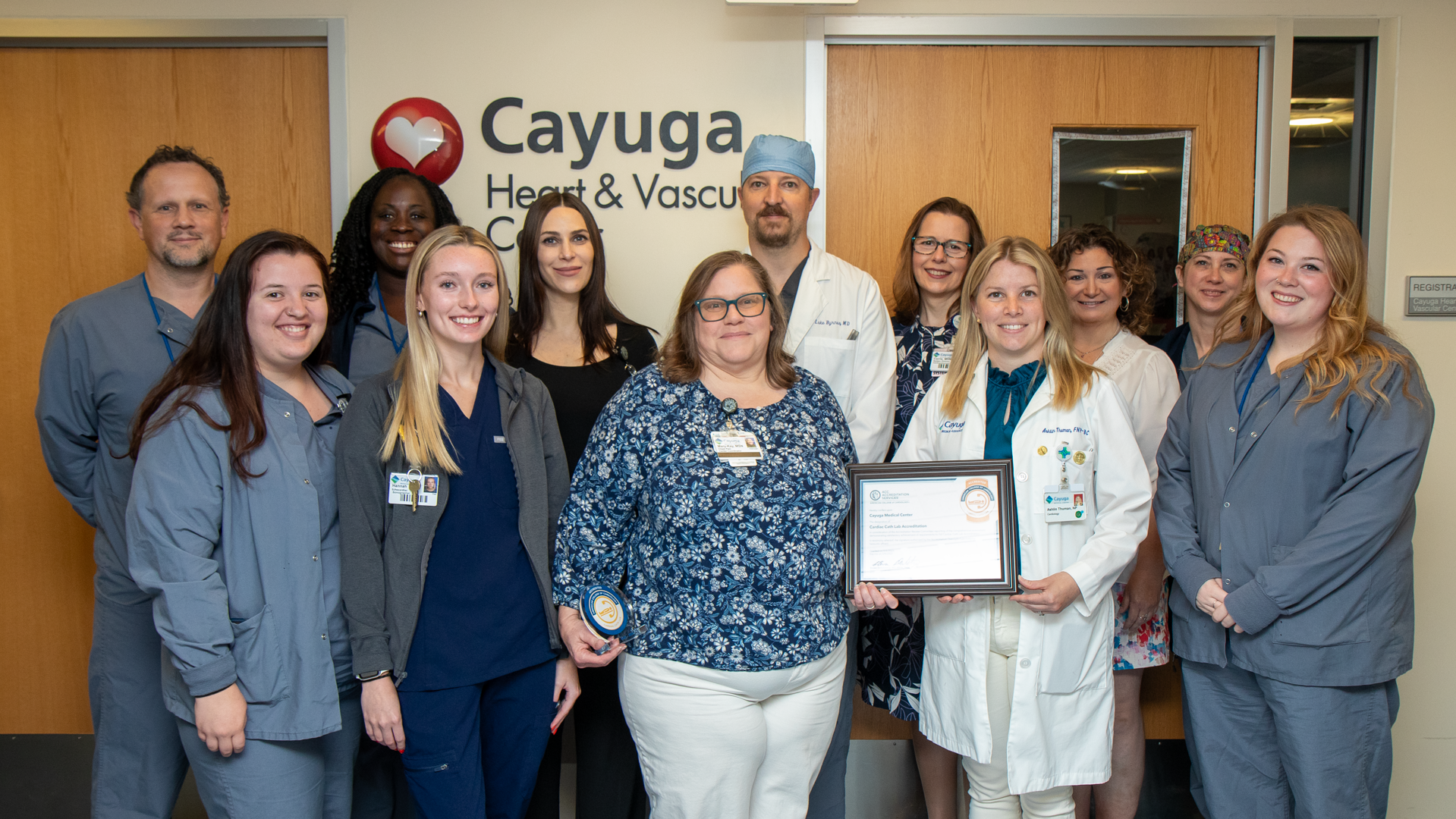
Cayuga Heart and Vascular Center has been providing cardiac services for forty years and has more than a decade of experience in invasive cardiac care.
Schedule and Appointment
Call today to schedule an appointment
Our Services
Cardiac Tests & Screening
Our cardiac care specialists provide a complete range of outpatient diagnostic and screening tests.
EKG (electrocardiogram) is a simple, painless test that measures how your heart is beating and whether your heart muscle has been damaged. At Cayuga Heart and Vascular Center we perform both adult and pediatric EKGs. More Information.
What can I expect during the test?
A cardiac technician will attach several small pads with wires (electrodes) to your chest, arms, and legs. These wires connect to a computer that records the activity of your heart and displays the heart’s electrical impulses on a screen. Your doctor can tell from the patterns how your heart is working. A copy of your EKG can be produced during the test, to be kept as part of your permanent medical record.
How and when will I get my test results?
The EKG will be read by a cardiologist or a physician credentialed in EKG interpretation. A computerized report will be provided to your primary care physician within 48 hours. Your doctor will review the results of the test with you.
Where is the test performed?
Electrocardiograms are performed at Cayuga Heart and Vascular Center at Cayuga Medical Center on the 3rd floor.
Echocardiogram is an ultrasound, or sonogram, of the heart. A sonogram uses sound waves to create images and is a safe, gentle, and fairly quick exam. An echo provides information on the anatomy and function of the heart. Measurements are taken of the walls and chambers to determine the size of the heart. The mechanical function, or strength of the muscle squeezing, is assessed. The valves are evaluated to make sure they are thin and free of calcification and that they are opening and closing well. The blood flow across the valves is measured, which provides information about the health of the valves, pressures within the heart and lungs, and functioning of the heart muscle. An echo is performed by a sonographer, a person who has been educated and trained to perform ultrasounds, and is interpreted by a cardiologist, a physician specializing in cardiac health and treatment. On occasion, the sonographer may request an echo enhancement agent to improve the quality of the study. The sonographer will partner with a registered nurse for intravenous site placement and enhancement agent administration.
How do I prepare?
There is no special preparation for this test. We suggest that you wear a two-piece outfit, as you will be asked to disrobe from the waist up. You will be given a gown to wear with openings so that the technician has access to your chest wall.
What can I expect during the test?
This test is performed by a sonographer with special credentials. You will be asked to lie on your left side. A hand-held instrument, called a transducer, is moved to various locations on the chest and upper abdomen. The transducer emits and receives high-frequency sound waves. Images of your heart muscle and valves will be displayed on a monitor. These images will also be recorded for further examination by a cardiologist. An echocardiogram typically takes about half an hour.
How and when will I get my test results?
The echocardiogram is read by a cardiologist. The test results are sent to your referring physician within 48 hours, who will review them with you.
Where is the test performed?
Echocardiograms are performed in the Cayuga Heart and Vascular Center at Cayuga Medical Center on the 3rd floor.
A bubble study is added onto some echocardiogram studies. It is used to evaluate whether or not there is communication (an opening) between the left and right sides of the heart. An IV is started and then we inject saline with tiny bubbles while you have a limited echocardiogram. The results of this test will be reviewed with you by the cardiologist the day of the test.
Transthoracic Echocardiogram is the most common type of echo. It is performed by a sonographer who uses a probe to acquire images of the heart and blood flow. The probe is placed on different locations on the chest to get various views of the heart. A small amount of gel is used to help acquire the images. EKG wires are placed on the chest to monitor the heart rate. The room is darkened for better visualization of the ultrasound images. The patient is positioned, typically on their left side, and may be asked to briefly hold their breath in or out to get better images. The exam takes approximately 30 to 60 minutes.
How do I prepare?
There is no special preparation for this test. We suggest that you wear a two-piece outfit, as you will be asked to disrobe from the waist up. You will be given a gown to wear with openings so that the technician has access to your chest wall.
What can I expect during the test?
This test is performed by a sonographer with special credentials. You will be asked to lie on your left side. A hand-held instrument, called a transducer, is moved in various locations on the chest and upper abdomen. The transducer emits and receives high-frequency sound waves. Images of your heart muscle and valves will be displayed on a monitor. These images will also be recorded on videotape for further examination by a cardiologist. An echocardiogram typically takes about half an hour.
How and when will I get my test results?
The echocardiogram is read by a cardiologist. The test results are sent to your referring physician within 48 hours, who will review them with you.
Where is the test performed?
Echocardiograms are performed in the Cayuga Heart and Vascular Center at Cayuga Medical Center on the 3rd floor.
Cardiac Stress Testing is used to evaluate the heart’s response to physical exercise and how well your heart handles the workload. As your body works harder during the test, it requires more fuel and your heart has to pump more blood. The test can show if there’s a lack of blood supply through the arteries that go to the heart. It helps your cardiologist determine if there are blockages or narrowing in the arteries in and around the heart. At Cayuga Heart and Vascular Center, we perform four types of cardiac stress testing: Exercise stress test, nuclear exercise stress test, nuclear pharmacological stress test, and exercise and pharmacological stress echocardiography.
How do I prepare?
Do not eat or drink for at least two hours before the test. Wear comfortable loose-fitting pants, top, and sneakers or walking shoes. If you are taking any medications, call your doctor ahead of time to find out if there are any special instructions you should follow regarding your medicine. Plan to be at the center for about two hours, including outpatient registration, equipment setup, and completion of the stress test.
What can I expect during the test?
You will be asked to remove your clothing from the waist up and you will be given a gown to wear during the test. A cardiac technician will attach several electrodes to your chest with adhesive, and an EKG will be obtained. You will then be asked to either walk on a treadmill, following protocols designed to make your heart work hard. While you exercise, your heart rate and blood pressure will be constantly monitored and the information stored in a computer for analysis and for comparison to your resting vital signs and EKG.
After the test?
You may resume your normal activities, though you might feel tired after this test, depending on your medical condition and your tolerance for vigorous exercise.
How and when will I get my test results?
The cardiologist will discuss the results with you immediately following the test and will also share them with your primary care physician.
Where is the test performed?
Cardiac stress tests are performed in the Cardiac Stress Testing Lab at Cayuga Medical Center, 101 Dates Drive, off NYS Route 96, in Ithaca.
Exercise Stress Testing involves walking on a treadmill or riding a stationary bicycle with electrodes attached to your chest. You are asked to follow protocols designed to make your heart work hard. While you exercise, your heart rate and blood pressure are constantly monitored and the information is stored in a computer for analysis and for comparison to your resting vital signs and EKG.
How do I prepare?
Do not eat or drink for at least two hours before the test. Wear comfortable loose-fitting pants, top, and sneakers or walking shoes. If you are taking any medications, call your doctor ahead of time to find out if there are any special instructions you should follow regarding your medicine. Plan to be at the center for about two hours, including outpatient registration, equipment setup, and completion of the stress test.
What can I expect during the test?
You will be asked to remove your clothing from the waist up and you will be given a gown to wear during the test. A cardiac technician will attach several electrodes to your chest with adhesive, and an EKG will be obtained. You will then be asked to either walk on a treadmill, following protocols designed to make your heart work hard. While you exercise, your heart rate and blood pressure will be constantly monitored and the information stored in a computer for analysis and for comparison to your resting vital signs and EKG.
After the test?
You may resume your normal activities, though you might feel tired after this test, depending on your medical condition and your tolerance for vigorous exercise.
How and when will I get my test results?
The cardiologist will discuss the results with you immediately following the test and will also share them with your primary care physician.
Where is the test performed?
Cardiac stress tests are performed in the Cardiac Stress Testing Lab at Cayuga Medical Center, 101 Dates Drive, off NYS Route 96, in Ithaca.
Nuclear Exercise Stress Testing uses radioactive materials called tracers to capture images of the heart functioning.
How do I prepare?
You may pre-register by phone one or two days prior to your test by calling the medical center at 274-4353. Avoid both caffeine and smoking for 24 hours before your test, and don’t eat or drink anything after midnight the night before. Bring all of your medications, in their prescription bottles, to the medical center on the morning of the test. Wear walking shoes and a comfortable two-piece outfit, and plan to be at the medical center for three to four hours. Because there are waiting periods, you may want to bring something to read. Be sure to let the technician know if you are diabetic, have chronic lung disease, suffer from knee or hip problems, or have had a stroke. Also alert the technologist to any drug or food allergies you have.
During the procedure
To get the heart muscle working hard, you may be asked to exercise on a treadmill for several minutes. When you cannot exercise any more, a tiny amount of tracer will be introduced into your body through an intravenous (IV) line in your arm. You will then be asked to lie very still for up to 30 minutes. As the tracer travels through your heart, a special camera (called a Gamma camera) detects the material and captures images of the blood flow in your heart muscle.
If you are unable to exercise you may be given a drug that will cause your heart to work as if you were exercising.
After the test
Most people can resume their normal activities after the test is completed. Talk with your doctor about when to take any medication you were asked to skip before the test. Your physician should be able to share the results of your test approximately 48 hours after its completion, and will provide a written report to your primary care physician.
Nuclear Pharmacological Stress Testing uses radioactive materials called tracers to capture images of the heart functioning. This test is much like nuclear exercise stress testing; however, this version of the nuclear cardiac stress test relies on a drug, rather than exercise, to get your heart muscle working hard.
How do I prepare?
You may pre-register by phone one or two days prior to your test by calling the medical center at 274-4353. Avoid both caffeine and smoking for 48 hours before your test, and don’t eat or drink anything after midnight the night before. Bring all of your medications, in their prescription bottles, to the medical center on the morning of the test. Wear walking shoes and a comfortable two-piece outfit, and plan to be at the medical center for three to four hours. Because there are waiting periods, you may want to bring something to read. Be sure to let the technician know if you are diabetic, have chronic lung disease, suffer from knee or hip problems, or have had a stroke. Also alert the technologist to any drug or food allergies you have.
During the procedure
A drug will be administered to you to get your heart muscle working hard, as if you were exercising vigorously. Once your heart is working hard, a tiny amount of tracer is introduced into your body through an intravenous (IV) line in your arm. As the tracer travels through your heart, a special camera detects the material and captures images of the blood flow in your heart muscle. You will then be asked to lie very still for up to 30 minutes. As the tracer travels through your heart, a special camera (called a Gamma camera) detects the material and captures images of the blood flow in your heart muscle.
After the test
Most people can resume their normal activities after the test is completed. Talk with your doctor about when to take any medication you were asked to skip before the test. Your physician should be able to share the results of your test approximately 48 hours after its completion, and will provide a written report to your primary care physician.
Exercise Stress Echocardiography uses echocardiogram high-frequency sound waves to capture images of the heart muscle at rest. You then walk on a treadmill or ride a stationary bicycle with electrodes attached to your chest. You are asked to follow protocols designed to make your heart work hard. While you exercise, your heart rate and blood pressure are constantly monitored and the information is stored in a computer for analysis and for comparison to your resting vital signs and EKG. At the end of exercising, your heart is scanned and these images are compared to the images of your resting heart to see if the muscle is functioning normally.
How do I prepare?
You may pre-register by phone one or two days prior to your test by calling the center at 274-4353. Avoid both caffeine and smoking for 48 hours before your test, and don’t eat or drink anything after midnight the night before. Bring all of your medications, in their prescription bottles, to the medical center on the morning of the test. Wear walking shoes and a comfortable two-piece outfit, and plan to be at the medical center for three to four hours. Because there are waiting periods, you may want to bring something to read. Be sure to let the technician know if you are diabetic, have chronic lung disease, suffer from knee or hip problems, or have had a stroke. Also alert the technologist to any drug or food allergies you have.
During the procedure
Cardiac nuclear medicine uses radioactive materials called tracers to capture images of the heart functioning. To get the heart muscle working hard, you may be asked to exercise on a treadmill or stationary bicycle for several minutes. When you cannot exercise any more, a tiny amount of tracer will be introduced into your body through an intravenous (IV) line in your arm. You will then be asked to lie very still for up to 30 minutes. As the tracer travels through your heart, a special camera (called a Gamma camera) detects the material and captures images of the blood flow in your heart muscle.
If you are unable to exercise you may be given a drug that will cause your heart to work as if you were exercising.
After the test
Most people can resume their normal activities after the test is completed. Talk with your doctor about when to take any medication you were asked to skip before the test. Your physician should be able to share the results of your test approximately 48 hours after its completion, and will provide a written report to your primary care physician.
Pharmacological Stress Echocardiography uses ultrasound (called an echocardiogram) to capture images of how the heart functions at rest. This test is much like the exercise stress echocardiogram testing; however, this version of the cardiac stress echo test relies on a drug, rather than exercise, to get your heart muscle working hard. Once your heart is working hard, the echocardiogram is used to capture images of the blood flow in your heart muscle. These images are stored and compared to the images of your heart muscle at rest, to look for abnormal movement or lack of movement in the heart muscle.
How do I prepare?
Avoid both caffeine and smoking for 48 hours before the test, and don’t eat or drink anything for four hours before the test. Wear comfortable loose-fitting pants, top, and sneakers or walking shoes. Bring all of your medications, in their prescription bottles, to the medical center on the morning of the test. If you are taking regular medications, call your doctor ahead of time to find out if there are any special instructions you should follow regarding your medicine. Plan to be at the medical center for about two hours, including outpatient registration, equipment setup, and completion of the stress echo.
What can I expect during the test?
You will be asked to remove your clothing from the waist up and you will be given a gown to wear during the test. A cardiac technician will attach several electrodes to your chest with adhesive, and an EKG will be obtained. Then a resting echocardiogram will be taken. You will be asked to lie on a bed on your back. Clear gel will be put on a transducer, which is a hand-held instrument that emits and receives high-frequency sound waves. The transducer will placed on two specific locations on your chest, and images of your heart muscle and valves will be displayed on a monitor. These images will also be recorded and stored in a computer.
The pharmacological cardiac stress echo test relies on a drug, rather than exercise, to get your heart muscle working hard. After the resting echocardiogram is completed, a drug (Dobutamine) will be slowly introduced into your bloodstream through an intravenous line in your arm. It will make your heart beat stronger and faster, causing it to work as it would during vigorous exercise. Your heart rate will be constantly monitored and the information stored. A second echocardiogram will be taken and the images of your heart will be compared to those taken prior to the Dobutamine, to look for abnormal movement or lack of movement in the heart muscle.
How and when will I get my test results?
The cardiologist will discuss the results with you immediately following the test and will also share them with your primary care physician.
After the test?
You may resume your normal activities, though you might feel tired after this test, depending on your medical condition and your tolerance for vigorous exercise.
Where is the test performed?
Stress echocardiograms are performed in the Cardiac Stress Testing Lab at Cayuga Medical Center, 101 Dates Drive, off NYS Route 96, in Ithaca.
PVD may affect any blood vessel outside of the heart. This includes the arteries, veins, or lymphatic vessels. Organs supplied by these vessels, such as the brain or legs, may not get enough blood flow for healthy function. The legs and feet are most often affected.
Peripheral vascular disease is also called peripheral arterial disease (PAD).
Diagnostic and Interventional Cardiology
Diagnostic and interventional cardiology refers to procedures that diagnose and treat coronary artery disease (including acute heart attack), certain heart valve problems, and congenital heart disease. These procedures are performed in the Cardiac Catheterization Suite and are minimally invasive because they rely on the use of catheters and tiny incisions rather than large incisions.
Diagnostic Cardiac Catheterization is an interventional procedure that enables your doctor to examine the coronary arteries and left ventricle function of your heart to diagnose a variety of potential heart problems. Your doctor may recommend this procedure if you are experiencing symptoms of coronary artery disease, such as chest pain, shortness of breath, or if other diagnostic tests such as an EKG or stress test indicate possible heart problems.
Your cardiologist will give you specific instructions prior to the catheterization regarding your medications. You may also be asked to have blood work performed at the medical center laboratory a week to ten days before the catheterization. Do not eat or drink anything after midnight the night before your cardiac catheterization. Because you will receive a mild sedative during the procedure, you must bring a family member or friend with you to drive you home afterwards. Cardiac catheterization patients typically return home the day of the test.
What can I expect during the test?
Cardiac catheterization is performed using local anesthesia. Either your wrist or leg is anesthetized, depending on where the catheter is inserted. You may also be given a mild sedative to help you relax. You are awake during the procedure since you may be asked to cough, breathe deeply, or hold your breath during the catheterization. The procedure itself is painless; however, you may feel warm or flushed for several seconds if a contrast medium is used.
How and when will I get my test results?
Your cardiologist will discuss your test results with you immediately following the cardiac catheterization. The findings will also be shared with your primary care physician. Based on this information, your doctors will be better able to diagnose your heart problem and advise you on your course of treatment.
After the test?
After the procedure you’ll be asked to lie quietly for a few hours. During that time, we will monitor your blood pressure frequently and inspect the access point regularly to be sure that all signs of bleeding have stopped. After discharge, you should limit your activities for one to two days and avoid any lifting.
Where is the test performed?
Cayuga Heart and Vascular Center at Cayuga Medical Center, 101 Dates Drive, off NYS Route 96, in Ithaca.
Percutaneous Coronary Intervention (PCI) is a procedure that uses balloon angioplasty and stents to open clogged coronary arteries on an emergency basis during a heart attack. PCI is also used electively in treating patients with chest pain due to coronary artery blockages. Balloon angioplasty initially opens the blockage and a tiny metal stent is then put in place to hold the blockage open while the artery heals.
Heart Rhythm Management
Holter Monitoring (ambulatory EKG) is a painless noninvasive method to record your heartbeat as you go about your daily activities.
How do I prepare?
There is no special preparation for this type of cardiac monitoring. A cardiac technician will give you guidelines to keep in mind during the monitoring period.
What can I expect during the test?
At the time you receive your Holter monitor, a cardiac technician will attach several small pads with wires (electrodes) to your chest. These wires connect to a small recording device that attaches to your belt. You will be asked to wear the monitor for a period of 24 hours or more. During that time you cannot shower, but a sponge bath is fine. Follow your normal daily routine, including work and exercise. Avoid using an electric blanket, as that can affect the recording. You will also be asked to keep a diary, noting times and activities, and any symptoms you feel.
How and when will I get my test results?
After you return the monitor, a cardiologist or EKG-credentialed physician will analyze the recorded data and prepare a written report for your primary care physician, who will discuss the results with you.
Where is the test performed?
Holter monitoring is available through the Cayuga Heart and Vascular Center at Cayuga Medical Center, 101 Dates Drive, off NYS Route 96, in Ithaca.
Cardioversion is a brief procedure during which a controlled electrical shock is delivered to the heart to convert or reset an abnormal heart rhythm to a normal rhythm. Elective or “non-emergency” cardioversion is most often performed to convert atrial fibrillation or atrial flutter.
Where is the procedure performed?
A cardioversion is done in the Cayuga Heart and Vascular Center of Cayuga Medical Center. It is scheduled for a minor procedure room where you will be monitored and prepared to receive moderate sedation.
How do I prepare?
Your physician will schedule the procedure and instruct you about which of your medications you should take beforehand. Take those medications on your regular schedule with a small amount of water. Otherwise you should not eat or drink anything for at least 8 hours before the procedure. Your physician may arrange for lab blood work to be drawn and processed prior to the cardioversion.
Please bring all of your medications with you to the appointment so we can verify your current medication dosages and schedules.
It will be unsafe for you to drive for the remainder of the day after receiving sedation, so please arrange for someone to provide you with a ride home.
What can I expect during the procedure?
When you arrive at the Outpatient Services area of the hospital, you will be greeted by a member of the cardiac services staff and escorted to the procedure room. At that time a brief review of your medical history will be done, along with baseline vital signs and any essential tests which need to be completed before hand.
You will be connected to a monitor that displays your heart beat, blood pressure, and oxygen level. Your cardiac nurse will start an IV (intravenous) line through which you will receive fluids and sedative medications for the procedure.
You will also be connected to the cardioverter machine. Two large patches will be applied to your chest to deliver the electrical shock for the actual cardioversion attempt. Oxygen is applied to your nose to help you breathe during the administration of sedation. The cardiologist is present in the room when everything is ready. She or he will actually coordinate the sedation and deliver the synchronized, controlled electrical shock to your chest wall for the cardioversion.
After the cardioversion has been done, a 12 lead EKG will be obtained to confirm return to normal sinus (regular) rhythm. You will be monitored for a period of time to ensure safe recovery from moderate sedation. You may experience some minor skin discomfort or irritation from the patches that were applied to your chest.
In some situations the actual cardioversion may be preceded by a transesophageal echocardiogram (TEE), a special ultrasound wave study (sonogram) to evaluate the structures and chambers of your heart. This is done to ensure that your heart chambers do not contain any problems which might complicate the safety of doing the cardioversion.
When can I go home?
After the cardioversion is complete we will monitor you for a period of time while you sleep off the effects of the sedation medications. When you are awake enough to control your swallowing, you may be allowed some fluids to drink. Once you are able to meet the criteria for safe recovery from sedation you will be discharged and may leave the medical center in the company of an adult relative or friend, who will drive you safely home. The recovery criteria include having a stable blood pressure, heart rate, and oxygen level; being awake and alert enough to be aware of your surroundings; and being able to sit up and stand without lightheadedness or dizziness.
Pacemaker Devices are very effective in treating people who have defective conduction systems in their hearts. Our hearts come with a natural pacemaker that regulates the rhythm of our heartbeat, slowing down when we’re at rest and appropriately speeding up when we’re active. Some people, however, develop faulty conduction systems in their hearts that prevent the rhythmic electrical impulses from traveling through the heart muscle. The result is often a heartbeat that is too slow or irregular. These conditions are very effectively treated with the implantation of a permanent pacemaker.
Pacemaker Implantation is an invasive procedure that takes place in the Cayuga Heart and Vascular Center. Patients receive a sedative and local anesthesia, which allow them to be mildly conscious during the procedure. The cardiologist makes a small incision in the shoulder and using guided imagery (a form of x-ray), attaches wires from the pacemaker to the inside of the heart muscle. The pacemaker is tucked beneath the skin on the shoulder.
Implantable Cardioverter Defibrillator (ICD) is a small device used to treat abnormal heart rhythms (called arrhythmias). ICDs are used in conjunction with proper medical therapy to treat patients who are at increased risk for sudden cardiac death from ventricular tachycardia or ventricular fibrillation. An ICD is a small computer that is surgically placed beneath the skin on the chest, with wires that attach to the inside of the heart muscle. This device monitors every heartbeat, detects arrhythmia, and delivers a shock to the heart. The shock disrupts the abnormal rhythm, with the goal of a restoring a normal heartbeat. ICD surgery is performed in the Cardiac Catheterization Suite at the Cayuga Heart and Vascular Center. Patients with ICDs receive lifetime management and support from our cardiologists and cardiac nurses.
Implantable Loop Recorder is a small device that is placed under the skin on the chest to help identify the causes of syncope (fainting). Syncope, which is a temporary loss of consciousness, may be caused by irregular heartbeats. The implantable loop recorder continuously records heart activity and can provide cardiologists with information about heart activity before, during, and after fainting episodes.
Wellness & Recovery
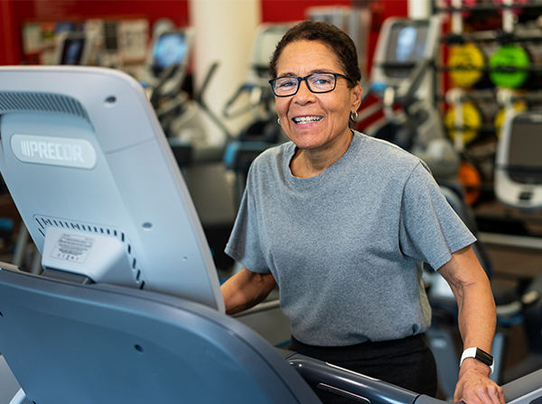
If you’ve had a heart attack or cardiac surgery, or if you suffer from stable angina (chest pain), your doctor may urge you to make lifestyle changes to improve your health and reduce your risk of future cardiac events. The care team at the Cayuga Center for Healthy Living (CCHL) located at the Cayuga Wellness Center, can answer your questions and guide you safely through recovery and into a healthier lifestyle. The Cardiovascular Disease Prevention and Cardiac Rehabilitation Program at Cayuga Center for Healthy Living (CCHL) has received program certification from the American Association of Cardiovascular and Pulmonary Rehabilitation.
Cardiovascular Rehabilitation through the Cayuga Center for Healthy Living works closely with the cardiologists at the Cayuga Heart and Vascular Center. One of the lifestyle changes your cardiologist is likely to recommend is regular exercise. The cardiovascular experts at CCHL can provide you with important answers about how you can feel safe exercising after a cardiac event, how much exercise is enough to be beneficial, and how much is too much.
With guidance and the direct supervision of our skilled team, you will learn more about your cardiovascular system, healthy eating, stress reduction, medications, the hows and whys of cardiovascular exercise, the limits imposed by your disease process, and the ways in which you can manage your individual risk factors for cardiovascular disease. Learn more about Cardiovascular Rehabilitation
ACCREDITATIONS
Accredited Chest Pain Center with Primary PCI and Resuscitation
![]() Hospital that have earned ACC Chest Pain Center with Primary PCI and Resuscitation Accreditation have proven exceptional competency in treating patient with heart attack symptoms and have primary PCI available 24/7 every day of the year. As required to meet the criteria of the accreditation designation, they comply with standard Chest Pain Center protocols and are equipped with a robust hypothermia program for post-cardiac treatment. These facilities also maintain a “No Diversion Policy” for out-of-hospital cardiac arrest patients.
Hospital that have earned ACC Chest Pain Center with Primary PCI and Resuscitation Accreditation have proven exceptional competency in treating patient with heart attack symptoms and have primary PCI available 24/7 every day of the year. As required to meet the criteria of the accreditation designation, they comply with standard Chest Pain Center protocols and are equipped with a robust hypothermia program for post-cardiac treatment. These facilities also maintain a “No Diversion Policy” for out-of-hospital cardiac arrest patients.
Hospitals receiving Chest Pain Center with Primary PCI and Resuscitation Accreditation from the ACC must take part in a multi-faceted clinical process that involves: completing a gap analysis; examining variances of care, developing an action plan; a rigorous onsite review; and monitoring for sustained success. Improved methods and strategies of caring for patients include streamlining processes, implementing of guidelines and standards, and adopting best practices in the care of patients experiencing the signs and symptoms of a heart attack. Facilities that achieve accreditation meet or exceed an array of stringent criteria. and have organized a team of doctors, nurses, clinicians, and other administrative staff that earnestly support the efforts leading to better patient education and improved patient outcomes.
Intersocietal Accreditation Commission (IAC)
![]() Certification by IAC is a very stringent process. IAC is the only accreditation board for echocardiography in the country and this certification confirms the high quality of our echocardiography capabilities. Cardiologists make critical decisions based on the anatomical and physiological information garnered from echocardiograms. The sonographers on staff at the Cayuga Heart Institute are highly experienced and use excellent technique, which is extremely important in the realm of cardiac care.
Certification by IAC is a very stringent process. IAC is the only accreditation board for echocardiography in the country and this certification confirms the high quality of our echocardiography capabilities. Cardiologists make critical decisions based on the anatomical and physiological information garnered from echocardiograms. The sonographers on staff at the Cayuga Heart Institute are highly experienced and use excellent technique, which is extremely important in the realm of cardiac care.
The National Cardiovascular Data Registry
 The National Cardiovascular Data Registry (NCDR) ® is one of the most comprehensive quality improvement programs in the country. NCDR tracks all data from the time a cardiac patient first enters a hospital’s care system and includes information on each patient’s symptoms, EKG and other test results, diagnosis, medications, additional chronic and acute illnesses (co-morbidities), and the specific medications and treatment provided to the patient in the hospital. NCDR and other mandated and voluntary registries provide state and national statistics on cardiac care trends, patient outcomes, and mortality. The information improves patient care by helping hospitals and doctors evaluate new equipment (such as coronary stents) and treatment methods. This enables us to refine our care accordingly to meet the highest current treatment standards.
ACR Accredited Nuclear Cardiology
 The Nuclear Medicine Department at Cayuga Medical Center is certified in Nuclear Cardiology by the American College of Radiology (ACR). Nuclear imaging is used to evaluate the function and anatomy of the heart while it is working and is a very important diagnostic test. ARC certification indicates that our nuclear cardiology services meet the highest standards of care.
AACVPR American Association of Cardiovascular and Pulmonary Rehabilitation
 The Cardiovascular Disease Prevention and Cardiac Rehabilitation Program at Cayuga Center for Healthy Living (CCHL) has received program certification from the American Association of Cardiovascular and Pulmonary Rehabilitation.
COLLABORATIONS
Pediatric Cardiology Associates of Syracuse
Rochester Heart Institute
Cayuga Medical Center’s Cayuga Heart Institute is a collaborator with the Rochester Regional Sands-Constellation Heart Institute (SCHI) in Rochester, New York. SCHI is a Cleveland Clinic Heart Surgery Center. US News and World Report has ranked the Cleveland Clinic Heart Surgery Center first in the nation in cardiac care for fifteen years running.
Our collaboration with the SCHI enriches local cardiac care by partnering Cayuga Heart Institute with a busy, progressive cardiac center noted for excellence in diagnosing, preventing, treating and managing heart disease and conditions. This relationship impacts our cardiac program in four distinct areas:
- Direct consultation on complicated cardiac patients, including the capability for immediate consultations during procedures, if needed.
- Quality review to ensure the highest possible standards of care, overseeing electrophysiology and interventional cardiology.
Driven by patient outcomes and in collaboration with the SCHI, the Cayuga Heart Institute has raised the bar in local cardiac care higher than ever. Our cardiac-care professionals strive to create a seamless system of care for patients from the moment they enter Cayuga Health System. Our care is patient focused, both on outcomes and on the way patients feel about how they have been treated.
For patients requiring interventional care beyond percutaneous coronary intervention (PCI) or cardiac catheterization, we provide a smooth transition to the finest, nearby cardiac center, the Sands-Constellation Heart Institute.
RESOURCES
Latest Heart News


Cayuga Medical Center Awarded Advanced Primary Stroke Center Certification from The Joint Commission


what our heart patients are saying

“As soon as Dr. Stefek for the last stent in, the pain totally disappeared”.

“They saved my life! They saved me from a major cardiac event and I am very thankful. Thank you for everything!”

“They have the doctors, staff, equipment, and experience to help you in a life or death situation like my heart attack.”